Technology

Technology

Technology

Technology

List of Technology and services we provide:
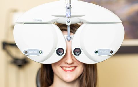
Marco Automated Phoropters
A Marco TRS 5100 automated phoropter is a state-of-the-art device used by our doctors during vision exams. More accurate diagnosis and better visual outcomes are the result of the precise and efficient measurements of a patient’s prescription with this device. Quickly switching between lenses, this phoropter makes the exam process faster and more comfortable. The TRS 5100 also eliminates the need for manual adjustments, reducing the likelihood of human error. Overall, this technology ensures the most accurate and up-to-date prescription for a patient’s glasses, ultimately improving vision and overall eye health.
Neurolens
Neurolens testing measures the way eyes work together. Using specialized equipment, eye movements will be tracked as the patient looks at a series of targets. This allows us to evaluate any potential misalignment or strain in the eyes which can cause headaches, neck pain, and eye fatigue. Should any issues be detected, neurolens glasses may be prescribed to help alleviate symptoms and improve visual comfort.
Learn More

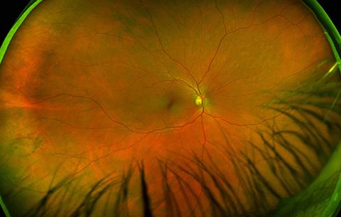
Optos Daytona Retinal Imaging
We offer digital retinal photography to all our patients. We use a 6.2 megapixel digital camera to photograph the blood vessels, macula, and optic nerve on the inside of the eye. This picture gives us an extremely accurate reference point to use in the future to monitor for changes such as diabetes, glaucoma, macular degeneration, and high blood pressure.
After the picture is taken, a photo comes up on the computer, and we can immediately review the results with you.
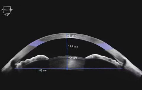
Zeiss Cirrus 5000 Optical Coherence Tomography (OCT)
Optical coherence tomography (OCT) is a non-invasive imaging test that uses light waves to produce high-resolution images of the eye's structures, both interior and on the surface. Through OCT imaging, eye conditions such as macular degeneration, glaucoma, and diabetic retinopathy can be diagnosed and monitored. The images produced by the OCT help to detect and track any changes to the structure of the eye.
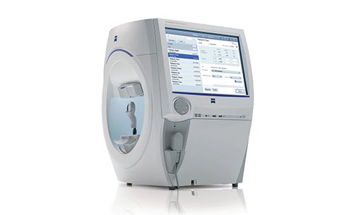
Zeiss HFA 3 Model 860 Visual Field Analyzer
Visual field testing is a procedure that measures the full extent of a patient’s peripheral vision. Critical in detecting eye conditions such as glaucoma, brain tumors, retinal detachment, and other visual impairments, this testing also helps to identify any abnormalities in vision and to monitor changes in field of vision over time. Early detection of eye diseases can prevent vision loss or blindness.
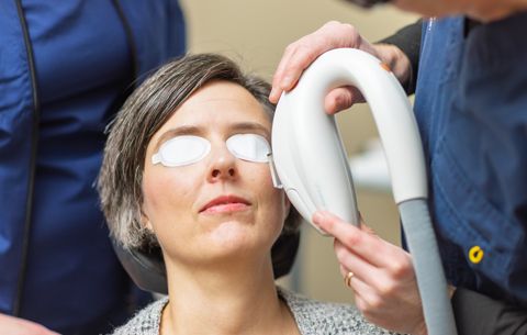
Lumenis Intense Pulsed Light (IPL)
Intense Pulsed Light is an in-office treatment targeting inflammation. Whether in the eyelids or on the skin surrounding the eyes, inflammation can exacerbate dry eye symptoms. IPL treatment has been shown to be effective in reducing dry eye symptoms, improving tear film stability, and increasing the production of oil from the meibomian glands in the eyelids. IPL is a non-invasive and well-tolerated procedure that can provide long-term relief for individuals with dry eye disease.
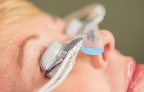
Lipiflow Thermal Pulsation
LipiFlow is an in-office treatment using thermal pulsation to unclog blocked meibomian glands in the eyelids These blocked glands cause dry eye disease. Taking about 12 minutes, Lipiflow involves placing a device on the eyelids that gently warms and massages the glands. This process helps to improve the quality of the oil produced by the glands, which can reduce dryness and irritation in the eyes.
Meibography
Meibography is a non-invasive imaging technique that allows us to see the structure and function of the tiny glands in the eyelids that produce the oil layer of tears. Using meibography helps us to diagnose and monitor conditions such as dry eye disease and to develop a personalized dry eye treatment plan.
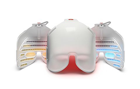
Equinox Low Level Light Therapy (LLLT)
Low-level light therapy (LLLT) is a non-invasive and painless treatment that involves exposing the eyelids and surrounding tissue to low levels of either blue light or red and near-infrared light. This therapy can improve dry eye symptoms by increasing blood circulation, reducing inflammation in the eyes, and increasing the oils in the tear film.
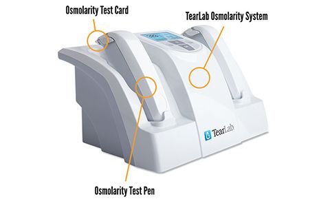
TearLab Tear Osmolarity Testing
Low-level light therapy (LLLT) is a non-invasive and painless treatment that involves exposing the eyelids and surrounding tissue to low levels of either blue light or red and near-infrared light. This therapy can improve dry eye symptoms by increasing blood circulation, reducing inflammation in the eyes, and increasing the oils in the tear film.
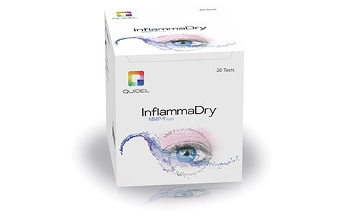
Inflammadry
Inflammadry is a simple and quick test used to detect inflammatory particles, which can be a part of dry eye disease, in tears. The test involves collecting a small sample of tears using a special device. The tears are then analyzed to measure the level of a specific protein that is present when there is inflammation in the eye. Results are available within minutes, allowing us to determine if further treatment is necessary to manage dry eye symptoms.
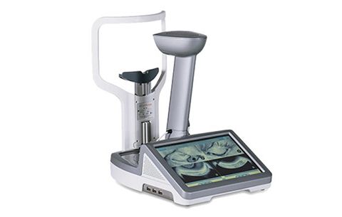
Oculus Keratograph 5M
The Oculus Keratograph 5M is a specialized device used to analyze the health and condition of the eyes' tear film and surface. Using a high-resolution camera and advanced software, the keratograph captures detailed images of the cornea, the transparent layer on the front of the eye, and measures the quality and quantity of tears.
At East Vancouver Eye, this technology is used to detect dry eye disease, meibomian gland dysfunction, and other issues affecting the surface of the eyes.


Powered by: